























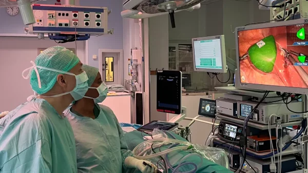
























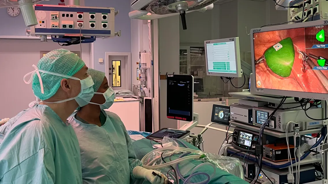
Gothenburg-based Navari Surgical, a Chalmers Ventures portfolio company, has used its augmented-reality surgical navigation technology on a human patient for the first time. The operation took place at Sahlgrenska University Hospital in June, and the company announced it on 19 October 2026.
The First Operation
Navari’s system is designed to help surgeons see tumours and other critical structures hidden beneath the surface of an organ. In the first operation the system worked as planned, the workflow ran well and the patient left hospital the day after surgery.
Axel Blomé, acting CEO of Navari Surgical, said: “Seeing the technology used on a human for the first time is incredibly big for us. It is a receipt for several years of hard work, but above all the beginning of the next phase. The potential of our technology goes far beyond liver surgery alone. Our vision is to give surgeons a completely new way of seeing and navigating the anatomy during an ongoing operation and, in time, make advanced navigation a natural part of modern surgery.”
Jonas Bergman, Investment Director at Chalmers Ventures, said: “Cancer surgery is an area with a great need for better tools. Navari addresses a concrete problem that surgeons around the world struggle with every day, and the fact that the technology has now been shown to work on humans is an important milestone.”
“A GPS for the Surgeon”
During an operation, an organ changes shape and position, so 3D images taken before surgery can quickly become less reliable. Getting a new ultrasound image normally means pausing the operation.
Navari’s system instead links the patient’s unique three-dimensional anatomy to the live surgical field. Using augmented reality, it shows tumours, vessels and other structures below the organ’s surface in relation to the surgeon’s camera view, updating continuously as the camera moves. Before surgery, the surgeon places a small marker with a QR code in the operating area; as long as the camera can see it, the system can show where the tumour and other key structures lie in depth.
The system also includes AI-supported calculations showing the position of surgical instruments relative to the tumour, and a complementary 3D view that gives the surgeon a perspective that a standard laparoscopic camera can’t.
Niclas Kvarnström, transplant surgeon and head of the Transplantation Centre at Sahlgrenska, said: “You can compare it to a GPS in the organ. Instead of just having a map before the operation, we get continuous guidance based on the current camera angle.”
From Clinical Need to Company
The work began about ten years ago after the opening of the Imaging and Intervention Centre at Sahlgrenska, when surgeons Niclas Kvarnström and Mårten Falkenberg began working with mathematicians Torbjörn Lundh and Klas Modin, professors at Chalmers and the University of Gothenburg. They studied how the surface of the liver could serve as a reference for locating deeper structures and developed a workflow for image support during surgery. The research led to scientific publications and several Chalmers master’s theses, and Navari Surgical was later formed to turn it into a medical device.
Lundh said: “This is a really good example of needs-based research that doesn’t start from what we’ve sat and thought up in peace at Chalmers, but instead has its basis in a real need from the operating room.”
The project has been funded by Vinnova, Chalmers Ventures and private investors, among others, and Region Västra Götaland has provided contract and innovation support.
What’s Next
Navari plans to use the technology in more operations this autumn, while continuing clinical validation, product development, quality systems and the regulatory process toward market approval. A larger international study is planned, and the company is already in talks with the interventional centre at Oslo University Hospital, among others. It will keep focusing on liver surgery, though use could widen over time to other organs and standard procedures. Navari’s ambition is to move from visualising anatomy to a broader platform for continuous surgical navigation.